

γ-Aminobutyric acid (GABA) is an important inhibitory neurotransmitter in the human central nervous system. In GABAergic neurons, when the presynaptic membrane is depolarized, the synaptic vesicles containing GABA will fuse to the presynaptic membrane and release GABA into the synaptic cleft. Subsequently, GABA binds to the GABAA receptor of the postsynaptic membrane, triggering Cl-influx, which hyperpolarizes the postsynaptic membrane and inhibits the transmission of nerve impulses. GABA transporter 1 (GAT1) is responsible for recycling GABA in the synaptic cleft to presynaptic membrane neurons, thereby regulating the activation state of downstream receptors. GAT1 maintains the stability of the neuroinhibited microcircuits, and its dysfunction can lead to a variety of neurological diseases, such as autism, epilepsy, and schizophrenia. GAT1 is an important anti-epileptic target, and by blocking the function of GAT1, it can inhibit the clearance of GABA in the synaptic cleft, thereby reducing nerve impulses to reduce epilepsy symptoms. In 1997, the US FDA approved the GAT1 inhibitor tiagabine as an adjunct to the treatment of epilepsy. Previously, the Gati group reported the electron microscopy structure of GAT1 bound to thiagabine at 3.8 angstroms in 2022 [1], but there is still a lack of high-resolution structural information to reveal the recognition and transport mechanism of GABA and the inhibition mechanism of other inhibitors such as 3-piperidinecarboxylic acid.
On July 3, 2023, the research group of Yan Chuangwei, associate professor of the School of Life Sciences of Tsinghua University, published important results on the transport and inhibition mechanism of GABA transporter GAT1 in Nature Structural & Molecular Biology. This work elucidates the mechanism of GAT1 transport substrate GABA through a combination of structural biology and biochemistry, reveals the inhibition mechanism of the competitive inhibitor 3-piperidinecarboxylic acid and the non-competitive inhibitor antiepileptic drug tiagabin, and lays the foundation for drugs that further target GATs.
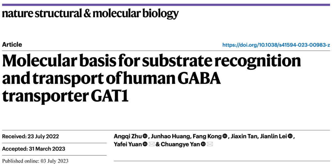
Figure 1 Structure of GAT1 in the substrate-free state and binding to three ligands
The analysis of low molecular weight membrane proteins is difficult in the field of cryo-EM, and generally requires the help of larger soluble regions or nanobodies. Previously, Yan's research group successfully resolved the high-resolution three-dimensional structure of the monocarboxylic acid transporter MCT1/Basigin-2 complex with flexible soluble region (3.0 angstroms) [2] and the glucose transporter GLUT4 (3.2 angstroms) [3] without soluble regions. In this work, the researchers further obtained a GAT1 structure with a high resolution of 2.2 to 2.4 angstroms without substrate binding and binding substrate GABA without nanobodies.
Thanks to the high-resolution electron microscopy density map, the researchers were able to accurately construct GABA, 3-piperidinecarboxylic acid, and tiagabine (Figure 2), while clearly observing sodium, chloride, and water molecules in the structure. The carboxyl groups of GABA and 3-piperidinecarboxylic acid can interact with sodium ions, residues G65 and Y140, while their nitrogen atoms can interact with surrounding water. By analyzing the interaction patterns of these ligands with GAT1, the study provides important insights into GAT1 recognition and transport substrates, as well as inhibition mechanisms. This research provides valuable design ideas for the further development of drugs for the treatment of neurological diseases targeting GATs.
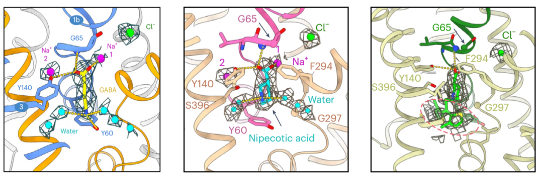
Fig. 2 Interaction patterns of GAT1 with GABA, 3-piperidinecarboxylic acid and tiagabin
(Note: This article is reproduced from the Internet)


 备案号:沪ICP备20003873号-1